Expert’s Desk · Knee
Knee cartilage lesion: what is it, and do you need surgery?

Short answer
A knee cartilage lesion is damage to a limited area of the smooth joint cartilage that covers the ends of the bones. It can range from mild surface softening to complete loss of cartilage down to the bone. Lesions are most often found on the back of the kneecap and on the inner joint surface of the thigh bone. Cartilage has a limited ability to repair itself. However, a cartilage lesion on an MRI does not by itself mean you need surgery: the same finding is seen in about 43% of adults aged 40 and over who have no knee symptoms and no history of injury.
Treatment is planned around the person, not the MRI picture. Symptoms, the size, depth and location of the lesion, any meniscus or ligament problem, leg alignment and sporting goals are weighed together. For lesions with few symptoms, the first step is usually strength exercise and load management. Surgical options range from arthroscopy that simply cleans up the cartilage to cell transplantation. That is why return to sport after "cartilage surgery" varies widely with the procedure: it is shorter after a clean-up alone, and averages 5 to 12 months after repairs that aim to grow new tissue. About four in five athletes return to sport after repair surgery, but getting back to the previous level is not guaranteed. If the knee locks, swells quickly after an injury or hurts too much to bear weight, see a doctor without delay.
Contents
- What is a cartilage lesion, and why does it develop?
- Cartilage lesions on an MRI report: what do the grades mean?
- Symptoms: how do you know it is a cartilage lesion?
- Non-surgical care: what is done, and what can you expect?
- Surgery: the options and the evidence
- Why do some people return in 2 months and others in a year?
- Recovery and return to sport: criteria, not the calendar
- Who tends to do less well?
- For clinicians
What is a cartilage lesion, and why does it develop?
Articular cartilage is the white, smooth tissue that covers the ends of the bones. It reduces friction during movement and spreads load across the joint surface. Because it has no blood supply, its capacity to heal after damage is limited. A cartilage lesion is damage confined to one area of this surface. It can begin as softening and surface cracks and progress to full-thickness loss of cartilage, or to damage that also involves the bone underneath (an osteochondral lesion).
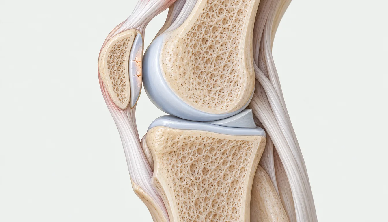
There are two main routes:
- Acute injury: a twisting knee, a kneecap dislocation, a direct blow or an ACL injury can damage the cartilage.
- Gradual damage: repetitive loading, kneecap tracking problems, loss of meniscus tissue or an unstable knee can wear the cartilage down over time.
A cartilage lesion is not the same as knee osteoarthritis. Osteoarthritis affects the joint as a whole, whereas a cartilage lesion is localized. Some lesions may, however, set the stage for osteoarthritis over time.
How common is it? In a study of 25,124 arthroscopies performed for knee complaints, cartilage lesions were found in 60% of patients. They were most common on the kneecap's joint surface (36%) and on the inner joint surface of the thigh bone (34%). The most frequent grade was partial-thickness damage. In 70% of lesions there was another problem as well, such as a meniscal tear or an ACL injury. These figures describe people whose knees were already troublesome enough to need arthroscopy, not the general population. A systematic review of studies in athletes reported an average prevalence of full-thickness cartilage lesions of 36%. That figure ranged from 2.4% to 75% between studies, and 14% of athletes with a lesion had no symptoms at the time of diagnosis.
Cartilage lesions on an MRI report: what do the grades mean?
MRI reports often give cartilage damage a grade. Softening and damage of the kneecap cartilage is often called chondromalacia or chondromalacia patella. Different classifications may be used in reports, but their logic is similar. The International Cartilage Repair Society (ICRS) grades are:
| Grade | What it means |
|---|---|
| 0 | Normal cartilage |
| 1 | Nearly normal: surface softening or very superficial cracks |
| 2 | Damage involving less than half of the cartilage thickness |
| 3 | Deep damage involving more than half of the thickness; may reach the bone but does not go through it |
| 4 | Damage passes through the cartilage into the bone beneath |
The grade on the report does not, on its own, show where the pain comes from. In a meta-analysis of 63 studies of adults with no knee symptoms and no history of injury, cartilage lesions were seen on MRI in 11% of people under 40 and in 43% of those aged 40 and over. MRI findings are therefore interpreted alongside the history and examination. We cover this in detail in My MRI report says there's a tear but I have no pain.
Symptoms: how do you know it is a cartilage lesion?
The symptoms of a cartilage lesion are often non-specific, meaning they resemble those of other knee problems:
- Knee pain under load, especially on stairs, when squatting or when standing up after sitting for a long time
- Recurrent swelling after activity
- Catching, or a gritty, grinding feeling inside the knee
- Locking, if a loose fragment of cartilage is floating in the joint
- Pain at the front of the knee with lesions behind the kneecap
These symptoms can also occur with a meniscal tear, with kneecap pain known as runner's knee, and in early osteoarthritis. That is why history, examination and imaging are interpreted together. A painless crack or click in the knee does not, on its own, mean cartilage damage; see our article on joint cracking. In athletes, unexplained recurrent knee swelling, painful or not, deserves careful assessment for a cartilage lesion.
MRI is the most useful test for showing cartilage. X-rays do not show cartilage directly, but may be used to assess leg alignment and signs of osteoarthritis.
Non-surgical care: what is done, and what can you expect?
For lesions with few symptoms, or those found incidentally on MRI, the first step is usually non-surgical care. Its main components are exercises that strengthen the front and back thigh muscles and the hip muscles, balance and movement-control work, conditioning, temporary adjustment of the loads that aggravate pain and, where relevant, weight loss.
There are no strong randomized trials comparing this approach directly with surgery. The available evidence comes from smaller studies and natural-history observations:
- An exercise programme: in Norway, 48 patients with full-thickness cartilage lesions followed a 3-month active rehabilitation programme of strength, neuromuscular and conditioning work. Average adherence was 83%, and knee function, muscle strength and hop tests improved meaningfully. Nine percent of patients could not continue the exercises because of pain and swelling. With no comparison group, these results are encouraging but not conclusive.
- Lesions left untreated: in one study, deep cartilage lesions found during ACL reconstruction were left alone; the menisci were intact and the lesions averaged 1.7 cm². An average of 8.7 years later, these patients' symptom scores were very close to those of a comparison group without cartilage lesions (about 93–94 vs 95–96 out of 100). In both groups, at least 79% of patients returned to jumping and pivoting sports at least at a recreational level.
- MRI follow-up: in Australia, the knees of 325 adults with an average age of 45, followed for research purposes, were scanned two years apart. Cartilage damage scores worsened in 33% of people and appeared to improve in 37%. Worsening was associated with female sex, older age and higher body mass index, while a fall in body mass index was associated with improvement. It is worth noting that these were not patients being followed for a particular injury.
These findings show that a cartilage lesion is not always a condition that inevitably gets worse. The course varies from person to person, and weight control and muscle strength are among the factors that may influence it. For general principles of load and exercise, our article on exercise for knee osteoarthritis may also help.
Surgery: the options and the evidence
Surgery is usually discussed when symptoms persist despite exercise and load management, when the lesion is of significant size and depth, or when a loose fragment is causing locking. The main procedures are:
| Procedure | What it involves | Usually for |
|---|---|---|
| Arthroscopic debridement | Loose, irregular cartilage edges are smoothed and any loose fragments removed | Small lesions, mechanical catching |
| Microfracture | Small holes are made in the bone at the base of the lesion to stimulate repair tissue | Small lesions; the repair tissue differs from the original cartilage |
| Osteochondral autograft transfer | Plugs of bone and cartilage from a less loaded part of the knee are moved into the lesion | Small to medium lesions |
| Autologous chondrocyte implantation | The person's own cartilage cells are grown in a laboratory and implanted in a second operation | Larger lesions |
| Osteochondral allograft | Donor bone-and-cartilage tissue from a tissue bank is used | Large lesions or those involving bone |
When a piece of bone and cartilage breaks off in an acute injury, such as a kneecap dislocation, it can sometimes be fixed back in place if it is suitable. This is a separate procedure and needs early assessment.
For small lesions, the simpler procedure holds its own. A double-blind randomized trial published in Norway in 2025 enrolled 65 patients aged 18 to 50. All had a lesion on the thigh bone smaller than 2 cm² (average 1.2 cm²) with no other knee problem. At two years, microfracture was not superior to arthroscopic debridement alone on any measure. Complications occurred 10 times in 5 patients after microfracture and twice in 2 patients after debridement. A review of debridement studies also reported good functional results in the short and medium term, while stressing that the level of evidence is low.
For large lesions, cell-based repair stands out in some trials. In a randomized trial of 144 patients with lesions of 3 cm² or larger (average 4.8 cm²), cell implantation improved pain and function more than microfracture at two years. Non-responders were 12.5% vs 31.9%, and the advantage in pain and function was maintained at five years. It should be noted that this trial was supported by the product's manufacturer. In the UK, a randomized trial followed 390 patients whose previous cartilage surgery had failed for 10 years (2026). Those who received cell implantation had knee function scores that averaged 7.3 points higher on a 100-point scale. However, treatment failure rates were similar in the two groups (29% vs 25%). When later operations were taken into account, the difference shrank and became statistically uncertain.
Caution is needed over the long term. In a randomized trial of 80 patients with a single lesion on the thigh bone treated with cell implantation or microfracture, there was no difference in clinical scores at 15 years. Treatment failed in 17 patients in the cell implantation group and 13 in the microfracture group. Among those without failure, 57% in the cell implantation group and 48% in the microfracture group showed early signs of osteoarthritis on X-ray. A network meta-analysis of randomized trials found similar reoperation rates and function scores across techniques at two years. Over the longer term, some advanced techniques had fewer reoperations.
The evidence can be summarized as follows: for small lesions, simpler procedures do not fall behind more complex ones. For large lesions, cell-based repair gives better function in some trials. However, no technique has been reliably shown to delay osteoarthritis.
Lesions behind the kneecap are a separate topic. A meta-analysis of repair surgery in this area (59 studies, 1,937 lesions) found clear improvement with most techniques. The overall failure rate was 6.8%, rising to 22.7% with donor grafts. About one in three patients had an anatomical risk factor related to kneecap alignment. That is why, for lesions in this area, how the kneecap tracks and the strength of the thigh and hip muscles are an important part of the treatment plan.
There is no strong evidence that injection treatments (PRP, hyaluronic acid, products marketed as stem cells) repair a localized cartilage lesion. For the broader evidence, see PRP and injection treatments.
Why do some people return in 2 months and others in a year?
"Cartilage surgery" is not the name of a single operation. When the cartilage is only cleaned up, or a loose fragment removed, no new tissue needs to mature, so return is usually quicker. With repairs that aim to grow new tissue, that tissue takes months to mature and return takes much longer. When the recovery times in the news seem not to add up, the reason is usually that different procedures were performed.
Key findings from studies of return to sport after repair surgery:
| Who, and which procedure? | Returned to sport | Average time to return |
|---|---|---|
| Osteochondral autograft transfer (meta-analysis of 44 studies) | 93% | About 5 months |
| Osteochondral allograft | 88% | About 10 months |
| Autologous chondrocyte implantation | 82% | About 12 months |
| Microfracture | 58% | About 9 months |
| All procedures, recent studies (52 studies, 2,387 people) | 80% | — |
| Professional athletes (11 studies, 476 athletes; mostly microfracture) | 84% | About 9 months |
Three points matter when reading these figures. First, most of the studies are of low evidence level. Second, the techniques are used for different lesions: osteochondral autograft transfer is used for smaller lesions, so part of its faster return may reflect the lesion itself. Third, returning to sport is not the same as returning to your previous level. In the recent review, only microfracture was associated with a tendency to return at a lower level. By sport, the likelihood of returning at the same level was higher in soccer and American football players and lowest in basketball players. Of 41 NBA players who had microfracture, 83% returned to professional basketball, and return to the NBA took an average of 9 months. However, players who returned appeared in fewer games per season and scored fewer points per game.
Recovery and return to sport: criteria, not the calendar
After repair surgery, rehabilitation usually begins with protected weight-bearing and range-of-motion work. It then moves on to progressive strengthening and, finally, to running, jumping and cutting. Experts recommend moving to each stage when specific criteria are met, not on a fixed date. Only a small share of studies in professional athletes report their return-to-sport criteria, which shows that this field is still developing.
Key criteria for returning to sport:
- No pain or swelling in daily life and during light running
- Full or near-full knee range of motion
- An acceptable strength difference between legs in the front and back thigh muscles
- Confidence and control in jumping and cutting tests
- No knee swelling the day after load is increased
For the general principles, see Return to sport: not the calendar, but the tests.
Who tends to do less well?
- People with large lesions or lesions involving bone: these are harder to repair and recovery may take longer.
- People with associated problems: a removed meniscus, ligament laxity, bow-legged or knock-kneed alignment, or kneecap tracking problems are addressed as part of the treatment plan. Otherwise, the repaired area keeps facing the same excess load.
- People with excess body weight: in the MRI follow-up study, a higher body mass index was associated with worsening lesions, and a fall in body mass index with improvement.
- People with several lesions or widespread osteoarthritis: here the principles of osteoarthritis care take priority over repairing one lesion.
- People who rush rehabilitation: increasing load quickly before the new tissue has matured may keep symptoms going.
This article summarizes the general evidence; it does not replace an examination by a doctor or an individual assessment.
For clinicians
Focal chondral and osteochondral defects are common, but prevalence data come from selected populations. Widuchowski et al. (n=25,124 arthroscopies) found chondral lesions in 60% of patients. Of documented lesions, 67% were localized focal lesions and 70% were non-isolated; the most frequent sites were the patella (36%) and MFC (34%), and the most frequent grade was Outerbridge II (42%). In Flanigan et al.'s systematic review of athletes (11 studies, n=931, all level 4), the prevalence of full-thickness focal defects was 36% (range 2.4–75%), with 14% asymptomatic at diagnosis. In asymptomatic, uninjured knees, MRI cartilage defects were present in 11% (95% CI 6–17) under 40 and 43% (29–57) at ≥40 years (Culvenor, BJSM 2019; 63 studies). Natural-history data are limited. In Ding et al. (n=325, mean age 45, 2.3 years), defect scores worsened in 33% of individuals and improved in 37%. Worsening was associated with female sex, age and BMI, and improvement with a fall in BMI (OR 1.23). This was not a cohort of focal traumatic lesions, and part of the score change may reflect measurement variability. In Shelbourne et al.'s series, untreated isolated Outerbridge 3–4 defects at ACL reconstruction (mean 1.7 cm², menisci intact) had subjective scores close to controls at a mean 8.7 years (94.0 vs 95.2 medial; 92.8 vs 95.9 lateral). There are no RCTs comparing non-operative care with surgery. In Wondrasch et al.'s 3-month active rehabilitation series (n=48, level 4), adherence was 83% and adverse events 9%, with significant improvement in IKDC, KOOS-QoL, isokinetic strength and hop tests. Surgery: in Randsborg et al.'s double-blind RCT (AJSM 2025; n=65, femoral, <2 cm², mean 1.2 cm²), MFX was not superior to arthroscopic debridement at 2 years (KOOS-QoL difference 3.5; 95% CI −10.0 to 16.9). Complications occurred 10 times in 5 MFX patients and twice in 2 debridement patients. In SUMMIT (Saris, AJSM 2014; n=144, ≥3 cm², mean 4.8 cm²; manufacturer-supported), MACI was superior to MFX for KOOS pain/function (P=.001), with non-responders 12.5% vs 31.9%; the difference was maintained at 5 years (Brittberg, AJSM 2018; P=.022). Knutsen et al. (JBJS 2016; n=80, first-generation ACI) found no clinical difference between ACI and MFX at 14–15 years. Failures were 17 vs 13, TKA 6 vs 3, and KL ≥2 in 57% vs 48% of survivors. ACTIVE (Snow, Osteoarthritis Cartilage 2026; n=390, prior failed surgery, mean 3.2 cm²) favoured ACI for 10-year Lysholm (mean difference 7.3; 95% CI 2.5–12.1). However, failure was 29% vs 25% (HR 1.04), and in the reoperation sensitivity analysis the difference fell to 3.8 (−1.8 to 9.5). Riboh's network meta-analysis (19 RCTs, n=855) found no difference in reoperation or Tegner/Lysholm at 2 years; at 5–10 years, OAT had lower reoperation than MFX and second-generation ACI lower than OAT. In the patellofemoral restoration meta-analysis (Hinckel, AJSM 2020; 59 studies, 1,937 lesions), overall failure was 6.8% and OCA failure 22.7%. The pooled rates of instability and anatomic risk factors were 11.9% and 32.1%, making alignment assessment essential. Return to sport: in Krych's meta-analysis (KSSTA 2017; 44 studies, n=2,549, level IV), the overall rate was 76% (OAT 93%, OCA 88%, ACI 82%, MFX 58%). Times were OAT 5.2±1.8 months, MFX 9.1±2.2, OCA 9.6±3.0 and ACI 11.8±3.8. In Kunze et al.'s contemporary review (AJSM 2025; 52 studies, n=2,387), the overall rate was 80.3%. MACI (OR 2.15) and OATS (OR 1.83) led for return at the same or higher level, MFX was associated with return at a lower level, and basketball had the lowest likelihood. In professional athletes (Kunze, Cartilage 2025; 11 studies, n=476, mostly MFX), the return rate was 84.3% at a mean 39.9 weeks; only 2 studies reported RTS criteria. In the NBA, return to professional basketball after MFX was 83%, mean time to return 9.2 months, with lower performance metrics (Harris 2013). Criteria-based progression is recommended in rehabilitation (Mithoefer, JOSPT 2012). There is no strong evidence that intra-articular injections achieve structural repair of focal defects.
Frequently asked questions
What is a knee cartilage lesion?
It is damage to a limited area of the smooth joint cartilage that covers the ends of the bones in the knee. It can range from mild surface softening to full-thickness loss of cartilage, or damage that extends into the bone beneath. Unlike osteoarthritis, it is confined to one area rather than affecting the whole joint. Seeing it on an MRI does not, on its own, mean it is the source of pain.
When does a knee cartilage lesion need surgery?
Surgery is usually discussed when symptoms persist despite exercise and load management, when the lesion is of significant size and depth, or when a loose fragment causes locking. Lesions found incidentally on MRI that cause no symptoms often do not need surgery. When surgery is needed, the options range from a simple arthroscopic clean-up to cell transplantation. In a randomized trial of small lesions, a simple clean-up did no worse than microfracture.
My MRI report says "grade 3–4 cartilage lesion". Do I need surgery?
Not on that basis alone. Grade 3–4 means the damage is deep or reaches the bone, but the decision depends on your symptoms and examination, not the MRI. Most patients whose deep lesions were found during ACL surgery and left untreated reported few symptoms even years later. The size and location of the lesion and any associated problems should be assessed with your doctor.
Can knee cartilage heal on its own?
Joint cartilage has a limited ability to repair itself because it has no blood supply. Even so, in adults followed with MRI, cartilage damage worsened over time in some people and improved in others. Managing body weight and keeping the leg muscles strong are among the factors that may influence this course.
How long after cartilage surgery can I return to sport?
It depends on the procedure. Return is quicker after a clean-up alone or removal of a loose fragment. After repair surgery, one meta-analysis found average return times of about 5 months after osteochondral autograft transfer, about 9 months after microfracture and about 12 months after chondrocyte implantation. These are averages; the actual decision is based on pain, swelling, strength and movement tests, not on the calendar.
What does a cartilage lesion behind the kneecap (chondromalacia) mean?
It is softening or damage of the cartilage on the back of the kneecap, where it faces the thigh bone, and it is one of the most common sites for cartilage lesions. It can cause pain at the front of the knee on stairs, when squatting or after sitting for a long time. Treatment focuses on strengthening the hip and thigh muscles and on kneecap alignment. This finding alone does not mean you have to stop sport; the decision is based on symptoms and examination.
Does a cartilage lesion turn into osteoarthritis?
It can, but not every lesion does. In patients followed for many years, some developed early signs of osteoarthritis on X-ray, whether or not they had surgery. No surgical technique has been reliably shown to delay osteoarthritis. Excess body weight, loss of meniscus tissue and poor leg alignment are factors that may increase this risk.
Related articles
My MRI report says there's a tear but I have no pain (or very little) — what does it mean?
KneeDoes exercise help knee osteoarthritis?
Return to SportReturn to sport after injury: not the calendar, but the tests
Scientific references
- Other references
- Widuchowski W et al. Articular cartilage defects: study of 25,124 knee arthroscopies. Knee 2007;14(3):177-182.
- Flanigan DC et al. Prevalence of chondral defects in athletes' knees: a systematic review. Med Sci Sports Exerc 2010;42(10):1795-1801.
- Culvenor AG et al. Prevalence of knee osteoarthritis features on magnetic resonance imaging in asymptomatic uninjured adults: a systematic review and meta-analysis. Br J Sports Med 2019;53(20):1268-1278.
- Brittberg M, Winalski CS. Evaluation of cartilage injuries and repair. J Bone Joint Surg Am 2003;85-A Suppl 2:58-69.
- Ding C et al. Natural history of knee cartilage defects and factors affecting change. Arch Intern Med 2006;166(6):651-658.
- Shelbourne KD et al. Outcome of untreated traumatic articular cartilage defects of the knee: a natural history study. J Bone Joint Surg Am 2003;85-A Suppl 2:8-16.
- Wondrasch B et al. The feasibility of a 3-month active rehabilitation program for patients with knee full-thickness articular cartilage lesions: the Oslo Cartilage Active Rehabilitation and Education Study. J Orthop Sports Phys Ther 2013;43(5):310-324.
- Randsborg PH et al. Microfracture versus arthroscopic debridement for the treatment of symptomatic cartilage lesions of the knee: 2-year results from a multicenter double-blinded randomized controlled trial. Am J Sports Med 2025;53(9):2107-2117.
- Totlis T et al. Arthroscopic debridement for focal articular cartilage lesions of the knee: a systematic review. Surgeon 2021;19(6):356-364.
- Saris D et al. Matrix-applied characterized autologous cultured chondrocytes versus microfracture: two-year follow-up of a prospective randomized trial. Am J Sports Med 2014;42(6):1384-1394.
- Brittberg M et al. Matrix-applied characterized autologous cultured chondrocytes versus microfracture: five-year follow-up of a prospective randomized trial. Am J Sports Med 2018;46(6):1343-1351.
- Knutsen G et al. A randomized multicenter trial comparing autologous chondrocyte implantation with microfracture: long-term follow-up at 14 to 15 years. J Bone Joint Surg Am 2016;98(16):1332-1339.
- Snow M et al. Ten year follow-up of a randomized trial of autologous chondrocyte implantation versus alternative forms of surgical cartilage management in the knee. Osteoarthritis Cartilage 2026;34(8):1243-1251.
- Riboh JC et al. Comparative efficacy of cartilage repair procedures in the knee: a network meta-analysis. Knee Surg Sports Traumatol Arthrosc 2017;25(12):3786-3799.
- Hinckel BB et al. Patellofemoral cartilage restoration: a systematic review and meta-analysis of clinical outcomes. Am J Sports Med 2020;48(7):1756-1772.
- Krych AJ et al. Return to sport after the surgical management of articular cartilage lesions in the knee: a meta-analysis. Knee Surg Sports Traumatol Arthrosc 2017;25(10):3186-3196.
- Kunze KN et al. High rate of return to sport for athletes undergoing articular cartilage restoration procedures for the knee: a systematic review of contemporary studies. Am J Sports Med 2025;53(10):2471-2482.
- Kunze KN et al. Return to sport in professional athletes after cartilage restoration surgery of the knee: a systematic review and meta-analysis demonstrates gender inequality and the need for improved reporting. Cartilage 2025;16(4):409-418.
- Harris JD et al. Return to sport and performance after microfracture in the knees of National Basketball Association players. Orthop J Sports Med 2013;1(6):2325967113512759.
- Thacher RR et al. Treatment of knee chondral defects in athletes. Sports Med Arthrosc Rev 2024;32(2):75-86.
- Mithoefer K et al. Current concepts for rehabilitation and return to sport after knee articular cartilage repair in the athlete. J Orthop Sports Phys Ther 2012;42(3):254-273.
This information is for general guidance only; it does not replace an examination, a diagnosis, or your physician's individual advice. Please consult a physician for your symptoms. About the author: Doç. Dr. Oğuz Yüksel — Academic Profile.